
Skin concerns · Uneven tone
Your skin is a palette of many tones.
The colour of your face comes from three sources: melanin, the skin's own pigment; the blood in your vessels; and the surface, which reflects light. When one of them stands out more than the others in a particular area, your tone looks uneven. Knowing which one dominates on your skin is the quickest way to the right care.
What the eye reads
The eye reads the spread of colour more than the colour itself.
Uneven tone shapes how rested, healthy or old a face looks. This finding sets no standard of beauty; it describes how the eye reads differences in colour as contrast. And most of the sources behind that contrast respond to care.
- 01
Age estimates
The facial colour distribution of 170 women aged 11 to 76 was transferred onto face models with identical shape and surface texture. These faces differed only in how their colour was distributed, yet the ages people gave them spanned roughly 20 years. Faces with an even tone were judged younger and healthier.[1]
- 02
Two pigments
In one study, 353 people rated cheek images of 170 women. The more even the tone, the younger the skin was judged to be. That evenness came down to the distribution of both melanin and haemoglobin, the pigment that gives blood its colour.[2]
- 03
Rosiness and health
When participants were asked to adjust face photos to look as healthy as possible, they increased the colour of oxygenated blood and gave the skin a slightly pinker cast. The issue is not red itself, but red that gathers in one area of the face.[3]
Differences in tone are very common and usually not a health concern. This information is here not to judge the face in the mirror, but to show you what can change.
Even spread
Patchy spread
The three sources of tone
Brown, red, sallow
Skin colour is largely set by three things: melanin and haemoglobin, which absorb part of the light, and the skin's tissue, which scatters it. Every difference in tone on your face comes down to one of these three, often to more than one.[2,4]
01
Brown
Melanin, the skin's pigment
Where: Epidermis, the upper layer of skin
Melanin is made by pigment cells in the upper layer of the skin and passed on to the cells around them. When sun, hormones or the inflammation left by a breakout step up production in one area, that area darkens. Marks left after breakouts and irritation appear on every skin tone; on deeper skin they are more frequent and more pronounced.[5]
Like the sun's ultraviolet rays, the blue-violet end of visible light can also trigger darkening. In a small study, this light caused darkening that lasted up to 3 months in skin types III and IV (medium skin that tans easily). Red light caused no darkening in the same study.[6]
What it looks like
- Sun spots
- Melasma
- Post-acne marks
- Freckles
02
Red
Haemoglobin, the colour of blood
Where: Dermis, the layer that holds the blood vessels
Red comes from the capillaries in the lower layer of the skin. When the vessels dilate, during frequent flushing or after irritation, it shows up as redness on the cheeks, nose and chin. If it fades for a moment when you press on it, the source is blood in the vessels.[7]
Blood does not only look red. Blood that collects beneath the skin can also read as a dark, patchy shadow; not every dark area is pigment.[4]
What it looks like
- Flushing
- Visible capillaries
- Irritation
- Rosacea
03
Sallow
Texture and light
Where: Stratum corneum, the outermost surface
No pigment gives this colour; it comes from the way the surface interacts with light. When proteins in the outermost layer oxidise, the surface lets less light through and turns opaque; as dead cells build up, light no longer reflects evenly. The result is a grey, dull look.[8]
A yellowish tone is also linked to glycation: sugar binds to proteins and advanced glycation end products (AGEs) accumulate. In a study of 40 women, cheek yellowness rose with both age and an AGE marker.[9,10]
What it looks like
- Dullness
- Yellowish tone
- Tired look
- Rough texture
Three steps in daylight
Map your own skin tone.
One morning, wash your face and stand at a window without makeup. Mark the colours you see in five zones; the map shows which source stands out and where to begin.
- 1
Light
Turn your face towards daylight. Bathroom lighting and screen light make tones look different from how they really are.
- 2
Colour
If you see a brown, red or sallow area that stands apart from its surroundings, mark it. A zone can have more than one colour.
- 3
Press
Press a red or dark area with your finger for a few seconds, then let go. If the colour fades for a moment, its source is blood in the vessels; if it stays the same, it is most likely pigment.[7]
| Zone | Brown | Red | Sallow |
|---|---|---|---|
| Forehead | |||
| Around the eyes | |||
| Nose | |||
| Cheeks | |||
| Chin |
You can also move through the table with the arrow keys.
Press test: does the coloured area fade for a moment when you press it?
Is there a mole or spot that has changed in size, shape or colour in recent months?
0 / 5 zones marked
Your tone map
You haven't marked a colour yet. If no zone clearly stands apart, your tone is largely even and your priority is protection: sunscreen every morning.
This map is not a diagnosis. The skin analysis in the app scores tone differences and under-eye darkness separately, so you can follow the change over the weeks.
Care by source
Each colour calls for different care.
Potent care aimed at brown tones can leave red skin redder still, while care that calms redness won't do enough for dullness. Choose the source first, then the care. Below, for each source, you'll find what the studies say and how strong the evidence is.
Brown
Melanin, the skin's pigment
- Every day
- Broad-spectrum sunscreen every morning. In a randomised study of 903 people, those who applied it daily showed no measurable increase in skin ageing over 4.5 years. If you are prone to melasma, choose a tinted (iron oxide) formula. In a study of 68 patients with melasma, both groups used the same dark-spot cream; in the group whose sunscreen also filtered visible light, the improvement in colour measurements was 28% greater at 8 weeks.[11,12]
- In your routine
- Vitamin C slows the work of tyrosinase, the enzyme involved in making melanin. In a study of 16 women with melasma, 5% vitamin C gave good or very good results in 62.5% of cases at 16 weeks. Hydroquinone, a dark-spot treatment, was stronger (93%), but side effects appeared in 1 of 16 people on the vitamin C side and in 11 on the hydroquinone side; colour measurements showed no significant difference between the two sides. Niacinamide acts at a different step, slowing the transfer of pigment to surrounding cells: in a study of 18 people, 5% niacinamide reduced dark spots compared with placebo within 4 weeks.[13,14,15]
- With Longos Sense
- Brightening: the stable vitamin C derivative 3-O-Ethyl Ascorbic Acid, with natural fruit acids from Kakadu plum and finger lime. Applied with the Quantum Light (red light), Radiofrequency and Cryotherapy modes of Longos Sense.
Evidence: Strong for sun protection; small but controlled studies for vitamin C and niacinamide.
Red
Haemoglobin, the colour of blood
- Every day
- Get to know your own triggers: heat, sun, alcohol, spicy food and harsh products can all increase redness. Wash your face with lukewarm water and pat it dry with a towel rather than rubbing.
- In your routine
- Simple, fragrance-free care that supports the barrier helps your skin react less. If redness persists in the centre of your face, a dermatologist is the right first stop: rosacea affects about 5.5% of adults and calls for medical care.[16]
- With Longos Sense
- Soothing: Tasmanian pepper extract and ceramide 3. Applied with the Cryotherapy mode of Longos Sense; cold temporarily narrows the small blood vessels in the skin.[17]
- Timeline
- The effect of cold is immediate and temporary. A calmer look comes with regular care and keeping away from your triggers.
Evidence: The vessel-narrowing effect of cold is well established; for persistent redness, it is no substitute for medical care.
Sallow
Texture and light
- Every day
- Sugar, smoking and sun speed up the build-up of glycation products (AGEs), which bind to proteins. Boiling and steaming instead of frying lower the amount of AGEs you take in from food.[9]
- In your routine
- Fruit acids (AHAs) loosen the bonds between dead cells and leave behind a surface that reflects light more evenly. Because they can make skin a little more sensitive to the sun, pair them with sunscreen during the day. And since oxidation turns the surface opaque, dull skin calls for sun protection and antioxidants together.[8,18]
- With Longos Sense
- Detox & Blue Light: extracts of Baikal skullcap root (Scutellaria baicalensis), cocoa seed and Epilobium. Applied with the Ultrasonic Waves mode of Longos Sense. The finger lime in Brightening also gently resurfaces the skin.
- Timeline
- As the surface renews, the first difference shows within a few weeks. Yellowness linked to glycation changes more slowly; here, protection comes first.
Evidence: Fruit acids have a long track record; findings on glycation and oxidation come mostly from laboratory and observational studies.
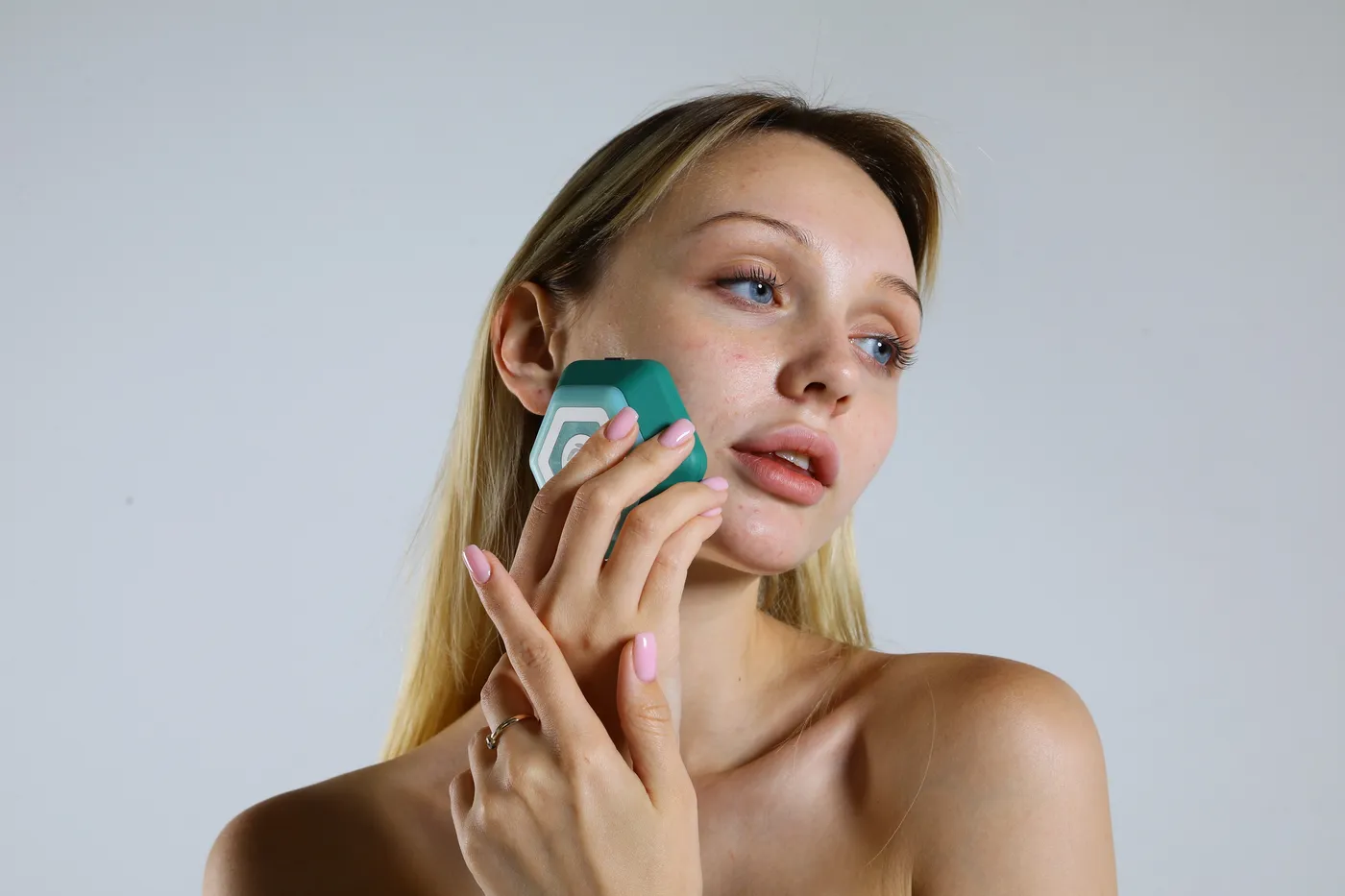
Longos Sense
One device, three modes for three sources
Longos Sense applies each serum in its own mode. Based on your skin analysis, the app sets which serum to use, in which mode and for how long.
For brown tonesBrightening
Red light Radiofrequency CryoFor red tonesSoothing
CryoFor sallow tonesDetox & Blue Light
Ultrasonic
Laboratory measurement
In the lab, the serums are more active with the device.[19]
Measured electrochemically (µA) in the laboratories of Yıldız Technical University and Dokuz Eylül University, with scientific contributions from AGON Biotechnology and Bioİzmir. This is a controlled laboratory observation, not a medical diagnosis or treatment claim.
All laboratory measurementsBrightening
+12%Increase in activity with the device
- Serum alone 19.6 µA
- With Longos Sense 22 µA
Soothing and Detox & Blue Light
4.5×Compared with Soothing measured alone
- Soothing alone 13 µA
- Detox & Blue Light alone 8 µA
- Both together, with Longos Sense 59 µA
Expert and user
A dermatologist and a user, in their own words.
The dermatologist on the Quantum Orbit Labs scientific board talks through the device and the skin analysis in the app; a user shares her experience over a few weeks, together with her analysis results.
Assoc. Prof. Dr. Sevil Savaş Erdoğan
Dermatologist · Interview on Longos Sense and skin analysis
Burcu Kara
Personal experience · Her analysis in the app after a few weeks: a more balanced skin tone, smoother texture
When to see a dermatologist
With these signs, see a doctor first.
Most differences in tone can be managed with care. Some signs, however, can be part of a medical condition; in that case, the right care starts after a doctor's assessment.
- 01
Redness that doesn't go away
Persistent redness in the centre of the face that flares from time to time; frequent flushing, visible capillaries, acne-like bumps or stinging eyes. These are among the diagnostic criteria for rosacea. Rosacea affects about 5.5% of adults; it is common and can be brought under control with medical care.[16,20]
- 02
A changing mole or spot
A spot that is asymmetrical, has an irregular border, more than one colour or is larger than 6 mm; above all, a mole that has recently changed in size, shape or colour, or that itches or bleeds. The ABCDE rule dermatologists use to assess moles lists these features; its most important letter is E, for evolving, meaning change.[21]
- 03
Dark patches that spread quickly
Brown patches that spread symmetrically across both sides of the face, whether during pregnancy, while taking hormones or over a short time, may be melasma. Melasma is harmless but stubborn; a dermatologist's opinion saves time in finding the right plan.
- 04
Yellowing all over the skin
If the whites of your eyes have turned yellow along with your skin, this is not a skincare matter. See a doctor without delay.
Frequently asked
About uneven skin tone
Is uneven skin tone the same as dark spots?
No. A dark spot is a clearly defined brown area and comes from just one source of tone: melanin. Uneven tone is broader, because redness on the cheeks and a dull surface also disrupt it. That is why care that works on dark spots may not be enough on its own for redness.
Which serum should I start with?
The colour you marked most on your tone map sets the starting point: Brightening for brown, Soothing for red, Detox & Blue Light for sallow tones. If more than one source is involved, the skin analysis in the app clarifies the order; the Signature Collection includes all three serums.
Can I use Brightening if my skin is red?
The vitamin C derivative and fruit acids in Brightening can sting sensitive skin. If redness stands out, calm your skin with Soothing first, then add Brightening on a few evenings a week, trying it on a small area to begin with. If stinging or redness continues, take a break and talk to your dermatologist.
Does red light darken dark spots?
In a small study on visible light, red light did not cause darkening; the darkening came from blue-violet light. Red light is part of the Longos Sense protocol for Brightening. Daytime sunscreen is still essential either way.
Does uneven tone look different on deeper skin?
The sources are the same, but they can look different. On deeper skin, redness may look purple or brownish rather than red, and the dark marks left after breakouts and irritation are more frequent and more pronounced. That is why gentle care that doesn't irritate matters especially on deeper skin tones.
When will I see a difference?
For redness, the effect of cold is immediate but temporary. For dullness, the first difference shows within a few weeks as the surface renews. In brown areas, pigment fades slowly; studies report results over months. Repeating the analysis in the app once a month, in the same light, lets you track the change with data.
References
Sources for the information on this page
Studies published in peer-reviewed journals, and laboratory measurements by Quantum Orbit Labs. The numbers in square brackets in the text link to this list.
- 1.Fink B, Grammer K, Matts PJ. Visible skin color distribution plays a role in the perception of age, attractiveness, and health in female faces. Evolution and Human Behavior. 2006;27(6):433-442. doi.org
- 2.Matts PJ, Fink B, Grammer K, Burquest M. Color homogeneity and visual perception of age, health, and attractiveness of female facial skin. Journal of the American Academy of Dermatology. 2007;57(6):977-984. pubmed.ncbi.nlm.nih.gov
- 3.Stephen ID, Coetzee V, Law Smith M, Perrett DI. Skin blood perfusion and oxygenation colour affect perceived human health. PLoS One. 2009;4(4):e5083. pmc.ncbi.nlm.nih.gov
- 4.Stamatas GN, Kollias N. Blood stasis contributions to the perception of skin pigmentation. Journal of Biomedical Optics. 2004;9(2):315-322. pubmed.ncbi.nlm.nih.gov
- 5.Davis EC, Callender VD. Postinflammatory hyperpigmentation: a review of the epidemiology, clinical features, and treatment options in skin of color. Journal of Clinical and Aesthetic Dermatology. 2010;3(7):20-31. pmc.ncbi.nlm.nih.gov
- 6.Duteil L, Cardot-Leccia N, Queille-Roussel C et al. Differences in visible light-induced pigmentation according to wavelengths: a clinical and histological study in comparison with UVB exposure. Pigment Cell & Melanoma Research. 2014;27(5):822-826. pubmed.ncbi.nlm.nih.gov
- 7.Sivakumar A, Thappa DM. Glass slide: an indispensable tool for the dermatologist. CosmoDerma. 2022;2:27. doi.org
- 8.Iwai I, Ikuta K, Murayama K, Hirao T. Change in optical properties of stratum corneum induced by protein carbonylation in vitro. International Journal of Cosmetic Science. 2008;30(1):41-46. pubmed.ncbi.nlm.nih.gov
- 9.Gkogkolou P, Böhm M. Advanced glycation end products: key players in skin aging? Dermato-Endocrinology. 2012;4(3):259-270. doi.org
- 10.Ohshima H, Oyobikawa M, Tada A et al. Melanin and facial skin fluorescence as markers of yellowish discoloration with aging. Skin Research and Technology. 2009;15(4):496-502. pubmed.ncbi.nlm.nih.gov
- 11.Hughes MCB, Williams GM, Baker P, Green AC. Sunscreen and prevention of skin aging: a randomized trial. Annals of Internal Medicine. 2013;158(11):781-790. pubmed.ncbi.nlm.nih.gov
- 12.Castanedo-Cazares JP, Hernandez-Blanco D, Carlos-Ortega B et al. Near-visible light and UV photoprotection in the treatment of melasma: a double-blind randomized trial. Photodermatology, Photoimmunology & Photomedicine. 2014;30(1):35-42. pubmed.ncbi.nlm.nih.gov
- 13.Pullar JM, Carr AC, Vissers MCM. The roles of vitamin C in skin health. Nutrients. 2017;9(8):866. pmc.ncbi.nlm.nih.gov
- 14.Espinal-Perez LE, Moncada B, Castanedo-Cazares JP. A double-blind randomized trial of 5% ascorbic acid vs. 4% hydroquinone in melasma. International Journal of Dermatology. 2004;43(8):604-607. pubmed.ncbi.nlm.nih.gov
- 15.Hakozaki T, Minwalla L, Zhuang J et al. The effect of niacinamide on reducing cutaneous pigmentation and suppression of melanosome transfer. British Journal of Dermatology. 2002;147(1):20-31. pubmed.ncbi.nlm.nih.gov
- 16.Gether L, Overgaard LK, Egeberg A, Thyssen JP. Incidence and prevalence of rosacea: a systematic review and meta-analysis. British Journal of Dermatology. 2018;179(2):282-289. pubmed.ncbi.nlm.nih.gov
- 17.Johnson JM, Kellogg DL Jr. Local thermal control of the human cutaneous circulation. Journal of Applied Physiology. 2010;109(4):1229-1238. pubmed.ncbi.nlm.nih.gov
- 18.Kornhauser A, Coelho SG, Hearing VJ. Applications of hydroxy acids: classification, mechanisms, and photoactivity. Clinical, Cosmetic and Investigational Dermatology. 2010;3:135-142. pubmed.ncbi.nlm.nih.gov
- 19.Quantum Orbit Labs. Electrochemical laboratory measurements of Longos Sense and the serums. Laboratories of Yıldız Technical University and Dokuz Eylül University; with contributions from AGON Biotechnology and Bioİzmir. quantumorbitlabs.com/en/clinical-evidence
- 20.Gallo RL, Granstein RD, Kang S et al. Standard classification and pathophysiology of rosacea: the 2017 update by the National Rosacea Society Expert Committee. Journal of the American Academy of Dermatology. 2018;78(1):148-155. pubmed.ncbi.nlm.nih.gov
- 21.Abbasi NR, Shaw HM, Rigel DS et al. Early diagnosis of cutaneous melanoma: revisiting the ABCD criteria. JAMA. 2004;292(22):2771-2776. pubmed.ncbi.nlm.nih.gov
Recommended products
Where to start for uneven tone
Longos Sense and three serums matched to the three sources of tone. Start with the serum for the colour you marked most on your tone map; the skin analysis in the app clarifies the order.
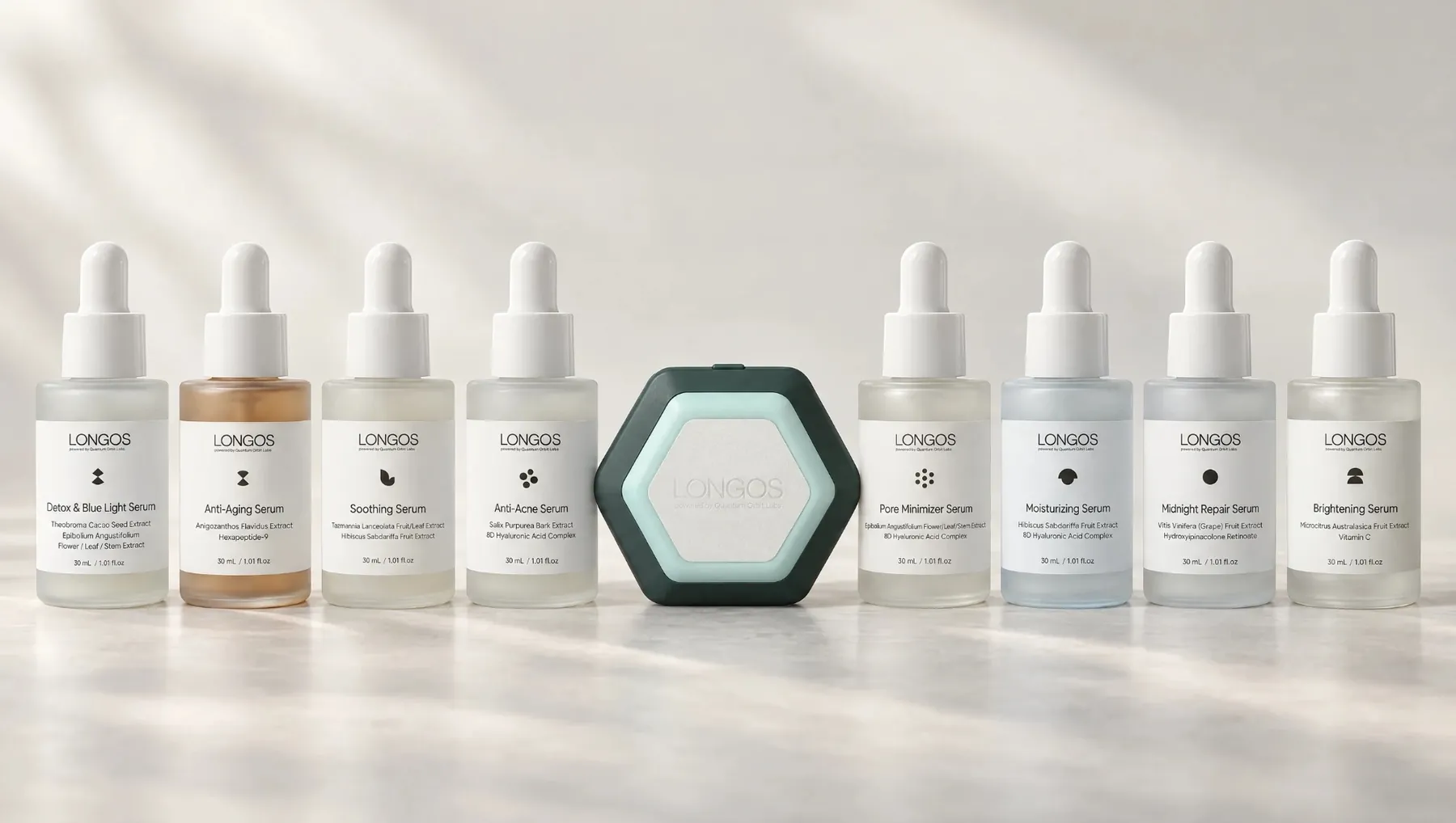
Longos Sense set
Quantum Light (red light), Radiofrequency, Cryotherapy and Ultrasonic Waves in one device. The app measures tone differences with a separate score and chooses the protocol; the set includes all three of these serums.
View the set-
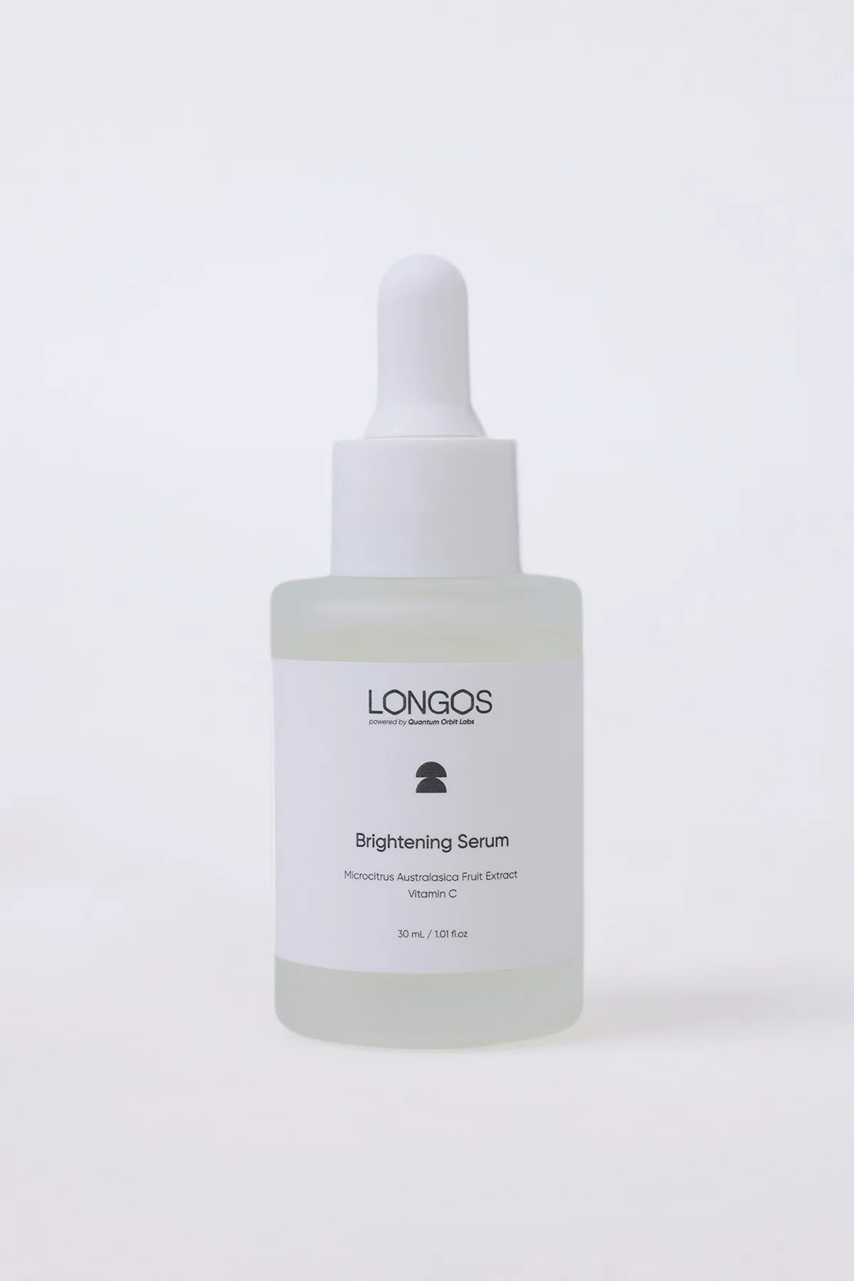
Brown · Melanin
Brightening serum
Stable vitamin C derivative, Kakadu plum and finger lime. Applied with red light, Radiofrequency and Cryotherapy.
Red light Radiofrequency Cryo -
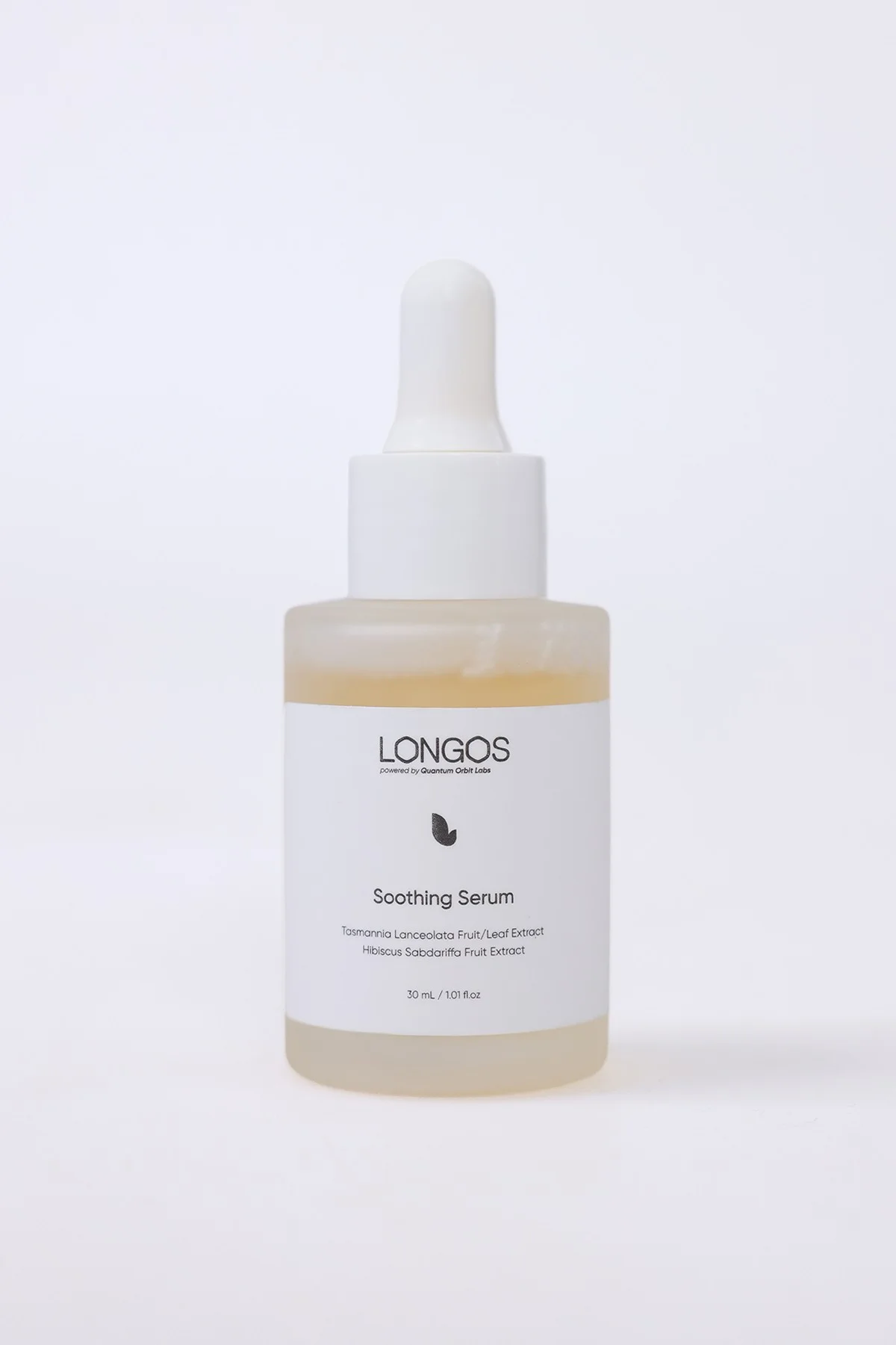
Red · Redness
Soothing serum
Tasmanian pepper extract and ceramide 3. Applied with the Cryotherapy mode on red, sensitive skin.
Cryo -
Sallow · Dullness
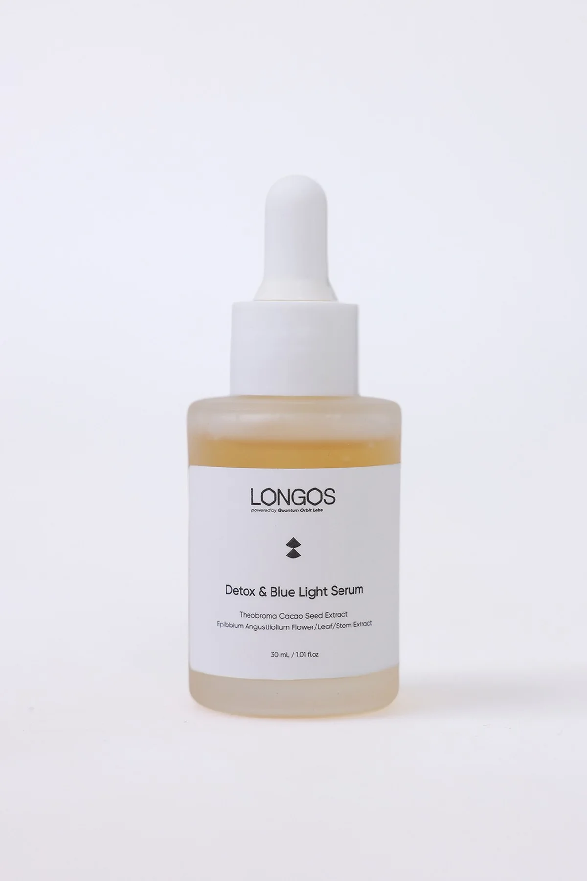
Detox & Blue Light serum
Extracts of Baikal skullcap root, cocoa seed and Epilobium. Applied with Ultrasonic Waves.
Ultrasonic
One morning this week, map your tone in daylight and run the skin analysis in the app the same day. Note your skin tone score; eight weeks later, repeat the analysis in the same light and compare the two scores.
Start your skin analysis